Medicine and Health
Osteoporosis cases found in many male smokers.

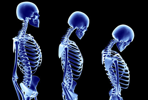
Osteoporosis is a disease that affects your bone mineral density by reducing it and by destroying the micro-architecture of the bone. It appears most frequently in female patients, particularly after menopause because of low levels of estrogen, which increase the mineral absorption from the bone into the blood stream thus reducing the bones density. Male cases are reported due to hormonal conditions or medications (glucocorticoids). This disease leads to numerous fractures mostly in the vertebral column, hips and wrists. Basically, your mineral density standard deviations are below average which produces some kind of bone fragility. It is important to add that osteoporosis fractures occur under some circumstances that normally don’t lead to fractures.
A recent large study consisted of middle-ageed to elderly smokers, concluded that smoking is also a risk factor for male cases of osteoporosis. Not only smoking but chronic obstructive pulmonary disease (COPD), are now recognized as risk factors for low bone density in both males and females. Guidelines don’t recommend osteoporosis screening for men because smoking history or COPD are not considered criterion for bone density screening. “Our findings suggest that current and past smokers of both genders should be screened for osteoporosis,” said Elizabeth Regan, MD, assistant professor of medicine at National Jewish Health. The National Jewish Health alongside other institutions have evaluated different types of ex-smokers with ages between 45 and 80 by using CT-scans to calculate their bone mass. More than 50% had low bone density and half of them also had one or more fractures of their vertebrae. Also, low bone density was increased in patients with severe COPD. They have evaluated the number of packs each participant smoked in pack-years of smoking and the average number was indeed higher in males with low bone density.
After all smoking is a high risk of osteoporosis even in men and also an additional risk to women. Considering the high cost that osteoporosis treatment has and the fact that it can only slow down the destructive process, osteoporosis screening for men should be considered in future guidelines.
Medicine and Health
A recently identified strain of deadly fungus poses a significant risk to public health
Researchers have recently discovered a new group of Candida auris, a potentially dangerous pathogen. The finding increases the total number of identified clades of the fungus, which is a newly emerging superbug resistant to multiple drugs, to six.
Candida auris is a strain of yeast that has the potential to cause serious illness and is frequently impervious to antifungal drugs. While individuals who are in good health generally do not fall ill, the transmission of the disease is highly prevalent within medical institutions and poses a significant risk to individuals with compromised immune systems. The yeast can induce a variety of conditions ranging from superficial infections of the skin to more severe and life-threatening illnesses, such as bloodstream infections. Due to its high level of resistance to multiple drugs, treating it can be challenging, and in some cases, even impossible.
The authors state that the pathogen is a significant global public health threat due to its widespread distribution, resistance to multiple drugs, high ability to spread, tendency to cause outbreaks, and high mortality rate. Although infections are still relatively uncommon, there has been a significant increase in cases in recent years.
Previously, the fungus had been categorized into five distinct clades, each located in different geographic regions: South Asia, East Asia, Africa, South America, and Iran.
In April 2023, doctors from the Singapore General Hospital identified a patient carrying a unique strain of C. auris as part of a routine screening program, adding it as the most recent clade to be discovered. Typically, these cases arise from individuals who have recently traveled, but this particular patient had not traveled outside the country for a period of two years, which raised some concerns.
Upon conducting a genetic analysis of the strain, the researchers determined that it did not align with any of the five known clades of the fungus. Therefore, it can be concluded that the strain belongs to a previously unidentified, sixth clade. Subsequently, they conducted tests on strains obtained from previous patients and identified two additional isolates of this particular group of C. auris in Singapore, as well as another isolate in Bangladesh.
The extent of the new clade’s prevalence and its potential to cause invasive infections and outbreaks remains uncertain at present. However, the researchers emphasize the importance of promptly identifying and controlling it in order to safeguard patient well-being.
“The ramifications of this breakthrough transcend the confines of the laboratory.” “Given the recent discovery of the sixth Candida auris clade, it is imperative to enhance surveillance capability or create new methods to strengthen existing surveillance strategies. This will enable health care facilities to closely monitor its emergence and effectively control its spread,” stated Dr. Karrie Ko, co-first author of the study.
Fortunately, the cases described in the study remained vulnerable to all antifungals that were tested. This should alleviate concerns about a pandemic similar to the one depicted in The Last Of Us. However, it is evident that the threat of C. auris is persistent. Therefore, additional efforts are required to identify new strains, monitor their spread, and control any negative clinical consequences.
The research is published in The Lancet Microbe journal.
Have you ever been scared so badly that you grabbed your chest? You feel like someone or something just zapped you behind the sternum. As you rest, you lean against the wall and think about why your friend is such a jerk and why you can feel it in your chest whenever you get scared.
People often use words like “heart-stopping” when they write fiction about fear, but the science of fear tells us that this isn’t what’s happening because it wouldn’t make sense. Our bodies are getting ready to deal with an impending threat when we’re scared, and going into cardiac arrest wouldn’t help us get very far if a lion was after us.
What do we do when we’re scared?
The sympathetic nervous system is what gets you excited when something makes you jump. It’s a tool inside our bodies that releases hormones and changes the way our bodies work to get us ready for the fight-or-flight response.
One important part is adrenaline, which is also known as epinephrine. The adrenal glands squeeze it out into the blood. The heart starts beating faster, sending more blood to your muscles and organs right away. Because they need all the oxygen they can get if they want to get away from a dangerous animal.
How do you feel when you go for a run?
Anyone who has ever used an EpiPen knows how bad it is to feel a sudden rush of adrenaline. It’s a stress hormone that makes you feel nervous and anxious, like you would before doing a bungee jump. Getting a rush when you think about a traumatic event from the past can be a sign of PTSD.
A medicine called adrenaline is used because it can help people who are having a medical emergency. If you have anaphylaxis from an allergen like peanuts, this can help because it can open your airway. Because it changes the strength and speed of heartbeats, it is also sometimes used to help people who are having a cardiac arrest.
When your adrenaline level goes up quickly, you may feel shaky, your heart beat quickly, and your chest get tight. When you add in the fact that you’re more alert, you become very aware of the changes in your body. This is especially clear when you’re not in danger, like when your partner surprised you at home when you thought you were alone.
When you’re scared, your sympathetic nervous system usually kicks in, which is normal. But, some heart conditions can get worse when you’re scared. Should anyone be having chest pain or ongoing discomfort, they should see a doctor. In the end, it is possible to be so scared that you die.
This article is not meant to be a replacement for medical advice, diagnosis, or treatment from a trained professional. If you have questions about a medical condition, you should always talk to a qualified health professional.
The family of Henrietta Lacks has filed a new lawsuit against two sizable drug companies for using her genetic material without her consent.
In the US District Court for the District of Maryland, Lacks’ living relatives are suing Novartis Pharmaceuticals Corporation, Novartis Gene Therapies, Inc., Viatris, Inc., and its subsidiary, Mylan Pharmaceuticals. They say the companies have used the “stolen” HeLa cell line to make hundreds of patents and have made a lot of money from it.
The suit wants the money made from using these cells to be “rightfully transferred” to Henrietta Lacks’s estate.
Novartis and Viatris chose to sell Henrietta Lacks’ living genetic material. Lacks was a black grandmother, community leader, and woman whose doctors took her tissue without her knowledge or permission, according to Chris Ayers, an attorney at Seeger Weiss LLP who is representing the Lacks family.
Ayers added, “We will keep looking for justice for Mrs. Lacks and her family.”
Henrietta Lacks died on October 4, 1951, from cervical cancer. She was 31 years old. Some of her cells are still alive today. A doctor at Johns Hopkins Hospital took a sample of her cervical cells without her knowledge just before she died. They were doing a cancer check. It was seen that her cells kept multiplying quickly, even after most of the cells in other samples would have died without their host.
Because scientists saw the potential, they found that these cells could be a cheap and easy way to help researchers do more research. The “HeLa immortal cell line” is what scientists call these cells, and they are very useful for biomedical research.
Over 75,000 scientific studies around the world have used these cells, which amount to about 55 million tons. They have been very important in making progress in areas like polio vaccines, cancer treatments, HIV/AIDS treatments, and much more.
All of this was done, though, without Lacks’ knowledge or permission. For many years, her family also didn’t know that the cells were being used for business.
Selling HeLa cells for money brings up important issues in medical ethics and genetics. As a black woman living in America in the 1950s, Lacks’ case shows how medical racism still affects minorities who aren’t getting enough help.
Even though a lot of people know about these problems, HeLa cells are still used in medical research for profit, which makes some companies a lot of money.
“Now that everyone knows Henrietta Lacks’ story, it’s shocking, but not surprising, that drug companies like Novartis and Viatris are still making money off of the deeply unethical origins of HeLa cells and the disturbing history of medical racism,” said Chris Seeger, another lawyer for the family.
A historic deal was made by Lacks’ family in 2023 after they sued Thermo Fisher Scientific, Inc., another biotech company, in the US District Court for the District of Baltimore. During that time, the lawyers said that the settlement was only the beginning and that there could be many more lawsuits about the use of HeLa cells.
- Gadgets10 years ago
Why the Nexus 7 is still a good tablet in 2015
- Mobile Devices10 years ago
Samsung Galaxy Note 4 vs Galaxy Note 5: is there room for improvement?
- Editorials10 years ago
Samsung Galaxy Note 4 – How bad updates prevent people from enjoying their phones
- Mobile Devices10 years ago
Nexus 5 2015 and Android M born to be together
- Gaming10 years ago
New Teaser For Five Nights At Freddy’s 4
- Mobile Devices10 years ago
Google not releasing Android M to Nexus 7
- Gadgets10 years ago
Moto G Android 5.0.2 Lollipop still has a memory leak bug
- Mobile Devices10 years ago
Nexus 7 2015: Huawei and Google changing the game